
|
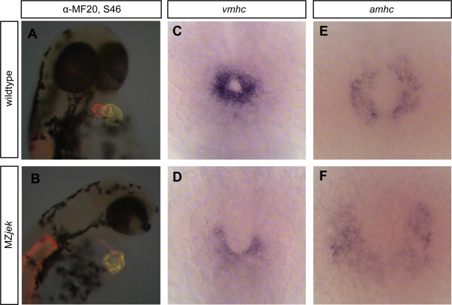
Fig. 2
MZjek have a dysmorphic heart and reduced ventricular populations. (A,B) Lateral views of hearts stained with MF20 (red) and S46 (green) antibodies to visualize the ventricle and atrium of 48 hpf embryos. MF20 marks the entire heart and S46 is atrium specific. In these superimposed images MF20+S46- ventricle is red and MF20+S46+ atrium appears yellow. (A) Wildtype heart with two distinct chambers. (B) MZjek display a dysmorphic heart, with a misshapen atrium and a strung out ventricle (N=9). (C–F) amhc and vmhc expression in wildtype (C,E) and MZjek (D,F) at the 20-somite stage. MZjek mutants exhibit reduced expression of vmhc when compared to similarly staged wildtype embryos (N=25, 36 respectively).
Reprinted from Developmental Biology, 387(2), Superina, S., Borovina, A., and Ciruna, B., Analysis of maternal-zygotic ugdh mutants reveals divergent roles for HSPGs in vertebrate embryogenesis and provides new insight into the initiation of left-right asymmetry, 154-166, Copyright (2014) with permission from Elsevier. Full text @ Dev. Biol.