
|
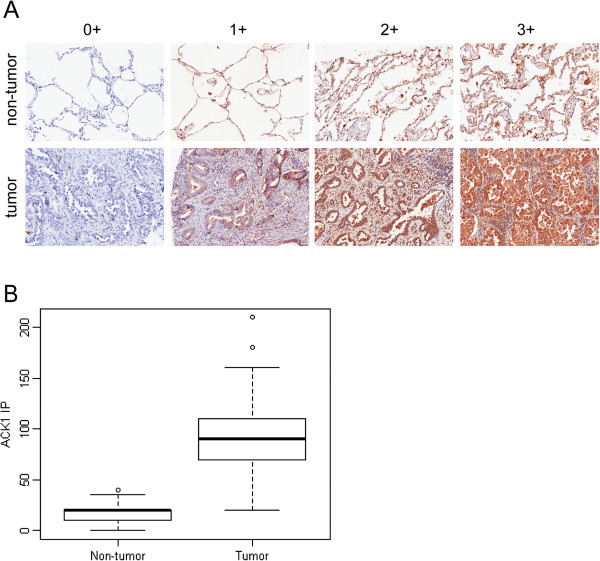
Fig. 5
ACK1 is highly expressed in lung adenocarcinoma. Immunohistochemistry staining was performed on 210 NSCLC tumor and paired non-tumor sections on an in-house TMA using anti-ACK1 (C20, see material and method). (A) ACK1 immuno-positivity was defined as presence of brown cytoplasmic staining. Staining intensity was scored as 0, 1+, 2+ and 3+ (no, weak, moderate and strong staining, respectively). (B) Percentage of positively stained tumor cells was assessed as proportion of total number of tumor cells present in the section. Intensity percentage score, IP, was defined as product of the maximum immunostaining intensity and percentage of tumor cells stained.