
|
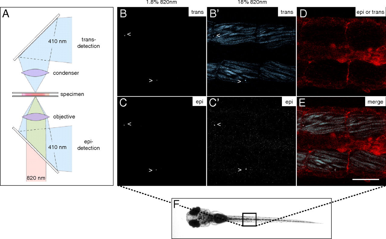
Fig. 4 SHG nanoprobes provide superior signal-to-noise ratio after in vivo injections BaTiO3 nanocrystals were injected into one-cell stage zebrafish embryos. Several days after cytoplasmic injection (around 72 hpf) excitation of nanocrystals with femtosecond pulsed 820-nm light results in strong SHG signal detectable in epidirection as well as in transdirection (A) throughout the whole zebrafish body [here, nanocrystals (arrowheads) present in the trunk of a zebrafish (B–C′)]. Endogenous SHG from trunk muscles can only be detected in transdirection with the sarcomere repeat patterns clearly observable (B′). (D) BODIPY TR methyl ester dye labeling the extracellular matrix and cell membranes. (E) Merge of pictures B′, C′, and D. Note that the power levels required to detect endogenous SHG are 10 times higher than those to visualize BaTiO3. The SHG signal intensity of BaTiO3 is comparable in epi- as well as in transdetection mode. Anterior to the left. Bar = 75 μm.