
|
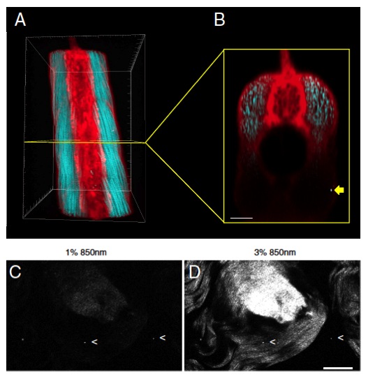
Fig. S4 SHG nanoprobes can be readily detected with increased imaging depth and provide superior signal-to-noise ratio when present in mammalian tail tendon. (A and B) BaTiO3 nanocrystals were injected into one-cell stage colorless zebrafish embryos. Several days after cytoplasmic injection (around 72 hpf) part of the zebrafish trunk was imaged with femtosecond pulsed 820-nm light. (A) Dorsal 3D reconstruction of the zebrafish trunk showing endogenous SHG signal from trunk-muscles detected in transdirection (blue), SHG of BaTiO3 nanocrystals detected in epi- as well as in transdetection (white), and BODIPY TR methyl ester dye labeling the extracellular matrix and cell membranes (red). Rostral front, caudal back. (B) Axial view of a slice of the trunk 3D reconstruction (yellow box). Whereas endogenous SHG and the vital dye are only readily detectable in the first 100 μm, the cross-sectional view through the trunk muscle shows that intense SHG signal of BaTiO3 nanocrystals is still detectable with increased imaging depth [here, nanocrystals (yellow arrow) present in the trunk of a zebrafish at an imaging depth of around 200 μm]. Note that the power levels required to detect endogenous SHG are 10 times higher than those to visualize BaTiO3. Bar corresponds to 50 μm. (C and D) BaTiO3 nanocrystals present in mouse tail slices of approximately 2–3-mm thickness. (C) One percent excitation with femtosecond pulsed 850-nm light results in strong SHG signal from diffraction-limited BaTiO3 nanocrystals (two of three spots marked with arrowheads) together with weak endogenous SHG signal originating from collagen in connective tissue of mouse tail tendon. (D) Increasing the excitation power level to 3% results in robust endogenous SHG signal with regular and highly organized collagen fiber clearly observable. Note that the power levels required to detect a strong signal from SHG nanocrystal signal spots is lower than for endogenous SHG of very highly aligned collagen fibers in mouse tail tendon. Bar corresponds to 50 μm.