
|
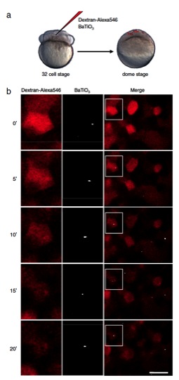
Fig. S3 SHG nanoprobes provide superior signal-to-noise ratio and do not bleach after in vivo injections. (A) Single-cell coinjections of BaTiO3 nanoparticles and 10,000 MW Dextran-Alexa546 were performed at the 32-cell stage and imaged at the dome stage of zebrafish embryos. (B) Time-lapse images of individual cells at the dome stage of a zebrafish embryo marked with BaTiO3 nanocrystals (Center column) and Dextran-Alexa546 (Left column). Right column is the merged picture with white boxes magnified in the center and left columns. Times (0′, 5′, 10′, 15′, and 20′) indicate minutes at and after the start of the experiment. Note that continuous scanning with intense excitation power levels bleaches quickly the organic dye, whereas the SHG signal of BaTiO3 stays unchanged, allowing long-term nanoparticle tracking. Anterior to the left. Bar corresponds to 20 μm.