
|
Fig. S2
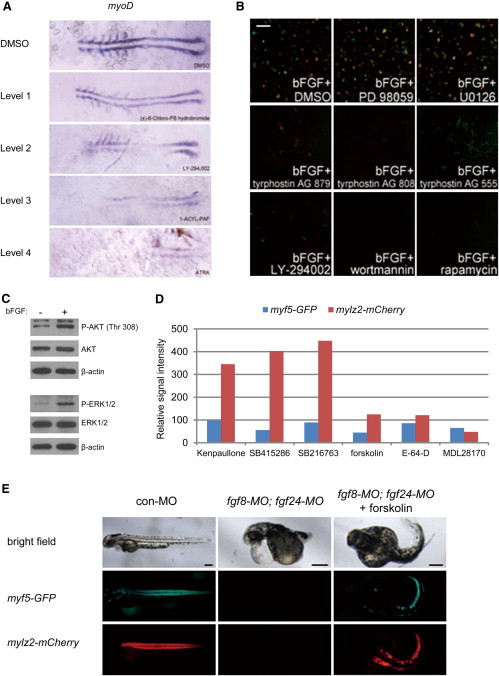
Chemical Genetics Screens to Identify Modifiers of Skeletal Muscle Development, Related to Figure 2
(A) Embryos were treated with chemical “hits” at the sphere stage. Once the control embryos reached the 6-somite stage, they were collected for whole-mount in situ hybridization with myoD riboprobe. Examples of hits verified to perturb muscle development in vivo. Flat mounted embryos stained with the myoD riboprobe at the 6-somite stage are shown with anterior to the left. Embryos were treated with (α)-6-Chloro-PB hydrobromide, LY-294002, 1-ACYL-PAF, or ATRA. All chemicals were from the CHB library and were diluted 300-fold. The level of myoD reduction was determined by comparing the intensity of myoD expression in these embryos with that in control embryos treated with DMSO.
(B) myf5-GFP;mylz2-mCherry embryos were collected at the oblong stage and dissociated. The resulting blastomere cells were aliquoted into a 96-well plate with pre-added chemicals. The culture medium contained 10 ng/ml bFGF to promote muscle development. Cells were treated with 50 μM tyrphostin AG 879, 50 μM tyrphostin AG 808, 50 μM tyrphostin AG 555, 50 μM PD 98059, 10 μM U0126, 10 μM LY-294002, 10 μM wortmannin, or 20 μM rapamycin. Cells were imaged after 2 days using a Celigo cytometer for GFP, mCherry, and bright-field signals. Scale bar represents 250 μm.
(C) bFGF treatment activates MEK/ERK and phosphoinositide-3-kinase (PI3K)-AKT signaling pathway in cultured blastomere cells. myf5-GFP;mylz2-mCherry embryos were collected at the oblong stage and dissociated. Resulting blastomere cells were cultured with or without bFGF. Cells were collected after 2 hr for Western blot.
(D) Hits from a screen without supplementation of bFGF in the culture medium. myf5-GFP and mylz2-mCherry signals were quantified by Image J and normalized to the DMSO-treated sample. Some inhibitors identified in this sensitized screen (e.g., SB216763) were missed in the initial screen, possibly due to toxicity or other unanticipated issues associated with the limited dose range that can be assayed in such a chemical genetic screen (see also Figure S6).
(E) 1-cell-stage myf5-GFP (green);mylz2-mCherry (red) embryos were injected with 250 pg fgf8-MO and 250 pg fgf24-MO and rescued by adding 10 μM forskolin at the dome stage. Scale bars represent 200 μm.
Reprinted from Cell, 155(4), Xu, C., Tabebordbar, M., Iovino, S., Ciarlo, C., Liu, J., Castiglioni, A., Price, E., Liu, M., Barton, E.R., Kahn, C.R., Wagers, A.J., and Zon, L.I., A Zebrafish Embryo Culture System Defines Factors that Promote Vertebrate Myogenesis across Species, 909-921, Copyright (2013) with permission from Elsevier. Full text @ Cell