
|
Fig. 2
Chemical Genetic Screens to Identify Modifiers of Skeletal Muscle Development
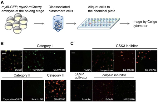
(A) Schematic of a high-throughput image-based chemical screening assay. Approximately 800 myf5-GFP;mylz2-mCherry double-transgenic embryos were collected and dissociated at the oblong stage. Resulting blastomere cells were aliquotted into four 384-well plates with preadded chemicals. After 2 days, the 384-well plates were imaged and analyzed using a Celigo cytometer.
(B) Sample images from the modifier screen. Hits could be grouped into three categories. Category I has only myf5-GFP expression. Category II has a decreased amount of both fluorescent colors. Category III has increased expression of both. Scale bars represent 250 μm.
(C) Hits from the enhancer screen. Six chemicals that increase the GFP and mCherry signals were identified. Scale bars represent 250 μM.
See also Figure S2 and Tables S1 and S2.
Reprinted from Cell, 155(4), Xu, C., Tabebordbar, M., Iovino, S., Ciarlo, C., Liu, J., Castiglioni, A., Price, E., Liu, M., Barton, E.R., Kahn, C.R., Wagers, A.J., and Zon, L.I., A Zebrafish Embryo Culture System Defines Factors that Promote Vertebrate Myogenesis across Species, 909-921, Copyright (2013) with permission from Elsevier. Full text @ Cell