
|
Fig. S1
Myogenesis Suppressors Identified In Vitro also Affect Muscle Development In Vivo, Related to Figure 1
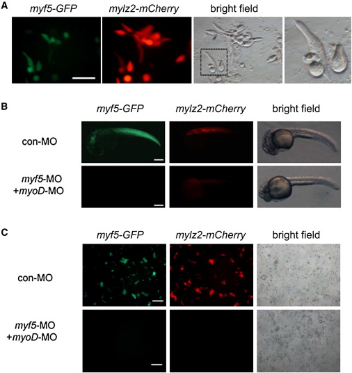
(A) Higher magnification of myf5-GFP;mylz2-mCherry blastomere cells cultured in zESC medium with bFGF. Scale bar represents 50 μm.
(B) myf5-GFP;mylz2-mCherry embryos injected with 400pg con-MO or co-injected with 200pg myf5-MO and 200pg myoD-MO lack both myf5-GFP and mylz2-mCherry expression. Images taken at 30 hpf. Scale bars represent 200 μm.
(C) myf5-GFP;mylz2-mCherry embryos injected with 400pg con-MO or co-injected with 200pg myf5-MO and 200pg myoD-MO were disassociated at the oblong stage and cultured in zESC medium with bFGF. No myf5-GFP or mylz2-mCherry expression was detected in the culture of double morphants. Images were taken 26 hr after plating. Scale bars represent 200 μm.
Reprinted from Cell, 155(4), Xu, C., Tabebordbar, M., Iovino, S., Ciarlo, C., Liu, J., Castiglioni, A., Price, E., Liu, M., Barton, E.R., Kahn, C.R., Wagers, A.J., and Zon, L.I., A Zebrafish Embryo Culture System Defines Factors that Promote Vertebrate Myogenesis across Species, 909-921, Copyright (2013) with permission from Elsevier. Full text @ Cell