
|
Fig. 1
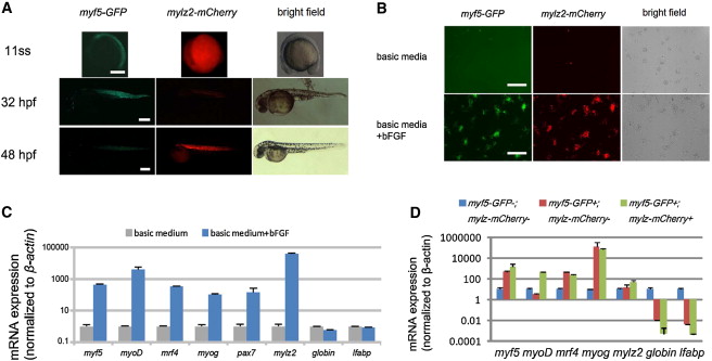
A Chemical Genetic Screen to Identify Modifiers of Skeletal Muscle Development
(A) myf5-GFP;mylz2-mCherry double-transgenic expression recapitulates expression of the endogenous genes. myf5-GFP is first detected at the 11-somite stage. mylz2-mCherry expression is not observed until 32 hpf. Scale bars represent 200 μm.
(B) myf5-GFP;mylz2-mCherry embryos were dissociated at the oblong stage and cultured in zESC medium. Images were taken 48 hr after plating. Scale bars represent 250 μm.
(C) Expression of myogenic genes measured by qRT-PCR. Blastomere cells were cultured and harvested at 24 hr. Error bars represent SD from the triplicate reactions. All values are normalized to β-actin and expression data of basic medium (gray bars, lacking bFGF) are set to 1.
(D) Expression of myogenic genes by different populations of blastomere cells isolated by FACS after 24 hr culture with bFGF. Error bars represent SD from triplicate reactions. Expression data are normalized to β-actin, and expression values of double-negative population (blue bars) are set to 1.
See also Figure S1.
Reprinted from Cell, 155(4), Xu, C., Tabebordbar, M., Iovino, S., Ciarlo, C., Liu, J., Castiglioni, A., Price, E., Liu, M., Barton, E.R., Kahn, C.R., Wagers, A.J., and Zon, L.I., A Zebrafish Embryo Culture System Defines Factors that Promote Vertebrate Myogenesis across Species, 909-921, Copyright (2013) with permission from Elsevier. Full text @ Cell