
Image
|
Figure Caption
Fig. S1
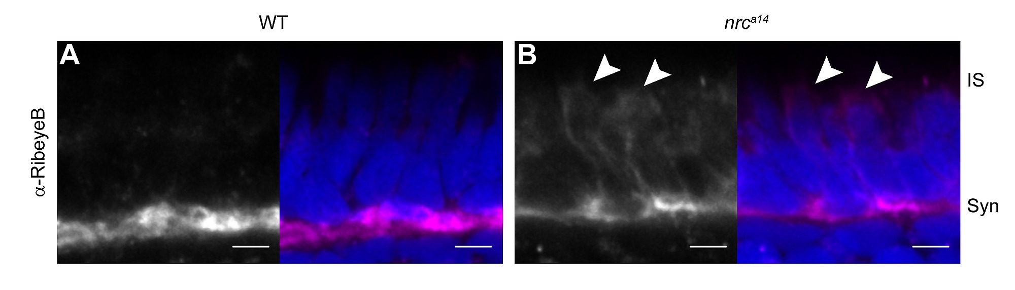
RibeyeB is mislocalized in nrca14 inner segments even in the absence of TαCP:spH. Anti-RibeyeB staining of non-transgenic WT and nrca14 5 dpf retinas showed the same staining pattern as Tg(TαCP:spH) 5 dpf retinas in Figure 2. In WT photoreceptors, RibeyeB was found only at synaptic terminals (A). In nrca14 cone photoreceptors, the RibeyeB staining was visible in both the synaptic terminals and ISs (B). Anti-RibeyeB staining is shown in magenta and Hoechst stained nuclei are in blue. Syn = photoreceptor synapses, IS = inner segment. Scale bar = 2 μm in all images.
Acknowledgments
This image is the copyrighted work of the attributed author or publisher, and
ZFIN has permission only to display this image to its users.
Additional permissions should be obtained from the applicable author or publisher of the image.
Full text @ PLoS One