
|
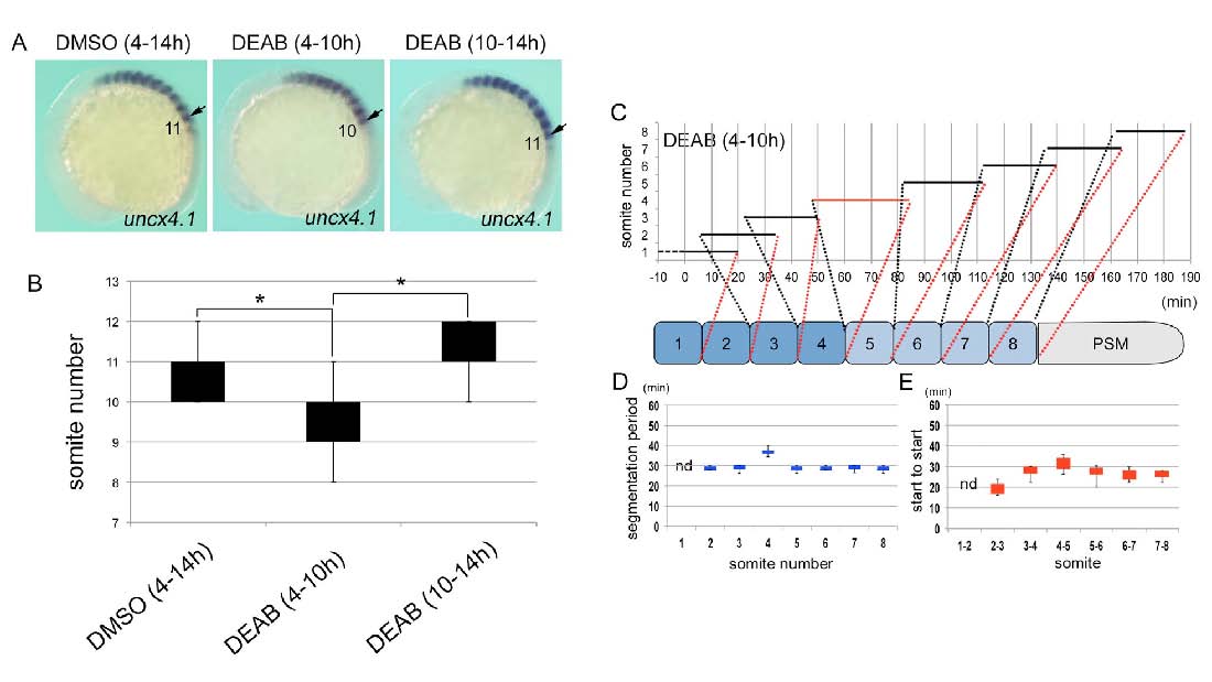
Fig. S6 Inhibition of RA synthesis during gastrulation results in loss of a somite. (A) Representative images of uncx4.1 expression in embryos treated with DMSO (4 – 14 hpf) (left), DEAB (4 – 10 hpf) (center) or DEAB (10 – 14 hpf) (right). Lateral view, anterior to the left. Arrows marked the position at SI. (B) Box and whisker plots of somite number in embryos treated with DMSO (4 – 14 hpf) (n = 34), DEAB (4 – 10 hpf) (n = 29) or DEAB (10 – 14 hpf) (n = 25). Statistically significant difference (asterisk, P < 0.05) could be seen in DMSO versus DEAB (4 – 10 hpf), and DEAB (4 – 10 hpf) versus DEAB (10 – 14 hpf). (C) Time-lapse data for somitogenesis in an embryo treated with DEAB (4 – 10 hpf) (see Movie 4, 4-1). (D, E) Box and whisker plots of segmentation period (D) or start to start (E) for embryos treated with DEAB during 4 – 10 hpf (n = 8). Results from statistical analyses are shown in Table S1 – 4.