
|
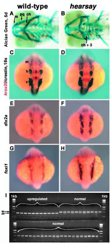
Fig. 8 Analysis of jaw and neural crest in hsy. (A,C,E,G) Wild-type and (B,D,F,H) hsy mutant embryos. (A,B) Alcian Green staining of cartilaginous jaw elements at 5 days. Ventral views, anterior towards the left. Cartilages: bh, basihyal; ch, ceratohyal; m, mandibular; pq, palatoquadrate; 3-7, gill cartilages derived from branchial arches P3- P7. m and pq are P1 derivatives; ch and bh are derived from P2. ch+3 indicates unilateral fusion of ceratohyal with gill arch 3 in mutant. (C-H) Expression analysis at the 18-somite stage: dorsal views, anterior towards the top. (C,D) Double labeling with crestin (dark purple) and krox 20 (red). (E,F) dlx2a expression. (G,H) foxi1 expression. (I) TaqI restriction polymorphism genotyping data for foxi1 in situ hybridization at the 18-somite stage, sorted as either ‘upregulated’ or ‘normal expression’. m, mandibular neural crest (nc) stream; h, hyoid nc; v, vagal nc; 3 and 5, krox20 expression in rhombomeres 3 and 5.