
|
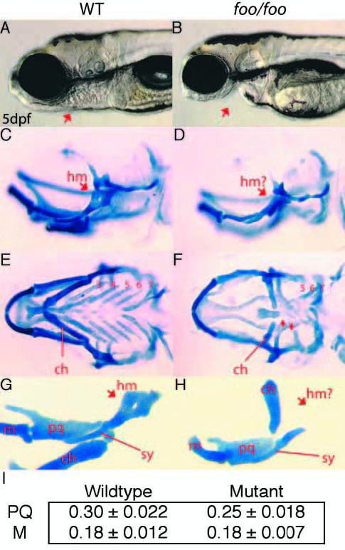
Fig. 5 foo mutants display defects in cartilages of the second, third and fourth pharyngeal arches. (A,B) Lateral views of wild-type (A) and foo/foo (B) embryos at 5 dpf. Arrow indicates jaw defects. (C,D) Lateral views of wild-type (C) and foo/foo (D) embryos at 5 dpf; wholemounts, Alcian Blue stained cranial cartilages. Arrow in D indicates the reduced hyomandibular region (hm) of the hyosymplectic. (E) Ventral view of C. The ceratohyal (ch) is indicated and gill arches are numbered 3-7. (F) Ventral view of D. The mildly reduced ch is indicated and arrows indicate the severely reduced third and fourth arch cartilages. The remaining gill arches 5- 7 are indicated. (G) Flatmount of animal in C. Arrow indicates the hm region; line indicates the symplectic rod region (sy). The palatoquadrate (pq), Meckel’s (m) and ch are also indicated. (H) Flatmount of animal in D. Arrow indicates the reduced hm while a line indicates the relatively normal sy. In foo/foo flatmounts, the ch positions abnormally because of an apparent defect in the articulation between ch and the hyosymplectic. (I) Chart comparing the measured length of the palatoquadrate (PQ) and Meckel’s (M) cartilage in wild-type and foo mutant embryos. Measurements were made of eight wild-type and six mutant embryos. The palatoquadrate shows a small but measurable reduction in size in foo mutants.