
|
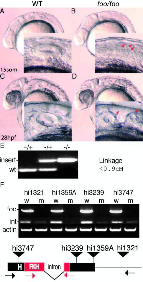
Fig. 1 Mutations in zebrafish foxi one cause defects in otic vesicle formation. (A) Lateral view of a wild-type embryo at 15 somites; inset is a magnification of the otic placode. (B) foo/foo embryo at 15 somites; arrows indicate putative placodes. (C) Wild-type ear at 28 hpf. (D) foo/foo embryo at 28 hpf; otic vesicle is clearly split into two smaller vesicles; arrows in inset indicate the two visible vesicles. (E) PCR products from genomic DNA indicating wild-type and mutant alleles of foo with the results of the linkage analysis in centimorgans. (F) RT-PCR analysis of each allele was performed. Wild-type, w; mutant, m. Bands marked foo use primers indicated by black arrows below; bands marked ‘int’ use primers indicated by red arrows. Integration for each allele is marked by a black triangle. The intron and the forkhead (FKH)-related DNA-binding domain are also indicated.