
|
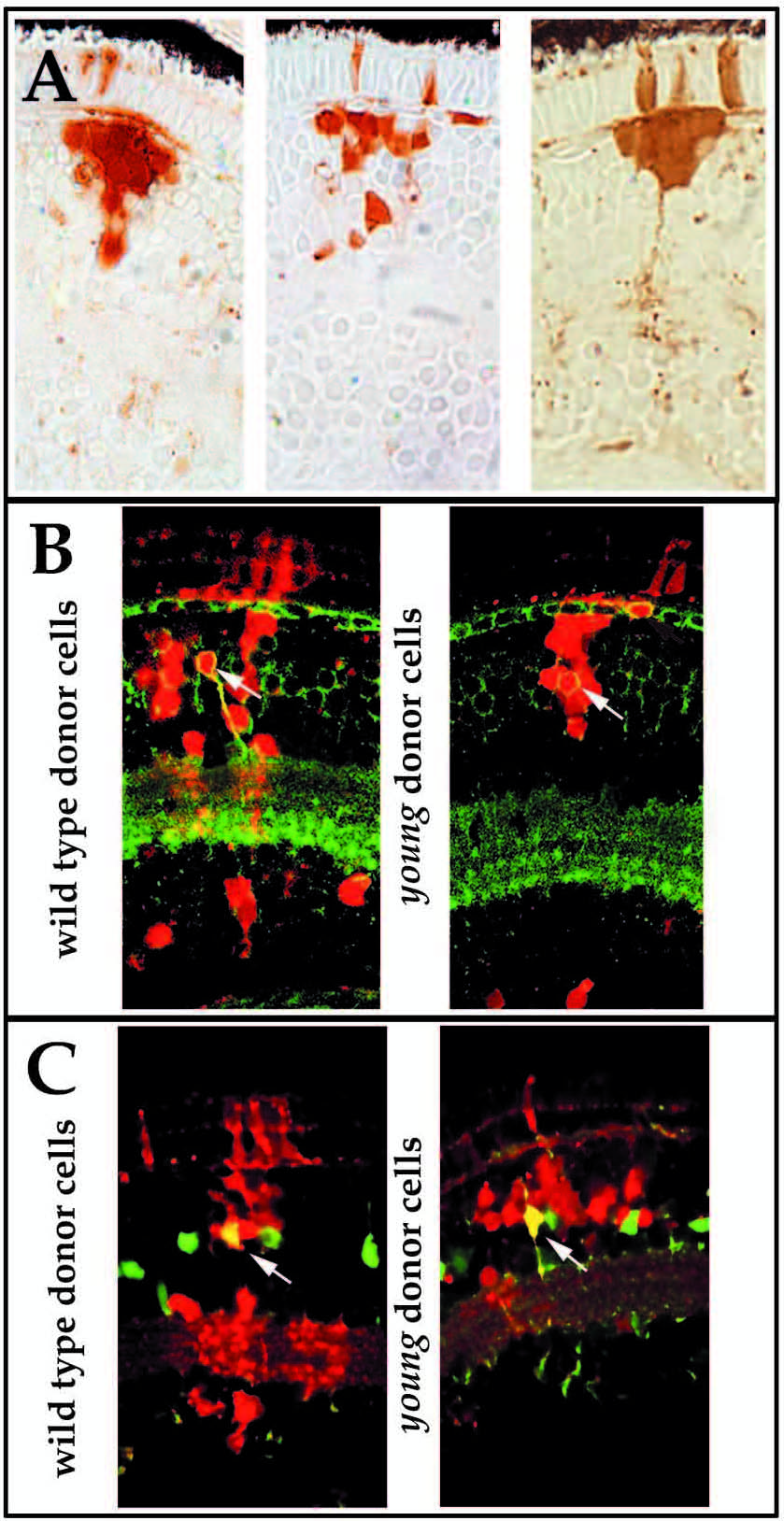
Fig. 5 Some young clones in wild-type hosts show a bias for cells of the outer portion of the inner nuclear layer. (A) Examples of three such clones at 3.5 dpf. (compare to clones of 4A). (B) Protein kinase C immunoreactivity marking bipolar cells and their processes in the inner plexiform layer and in close apposition to horizontal cells and photoreceptor terminals in the outer plexiform layer (green) in wildtype (left) and young (right) derived clones (red) at 8 dpf. (C) Carbonic anhydrase II immunoreactivity marking Muller glial cells in wild-type (left) and young (right) derived clones (red) at 5 dpf. Co-localization of molecular markers with young donor cells (yellow cells indicated with arrows) demonstrates that biased mutant clones contain both bipolar and Muller glial cells.