
|
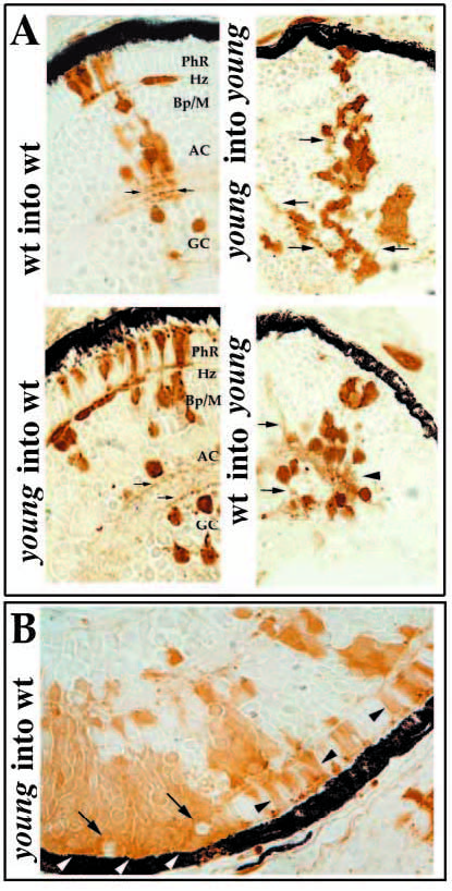
Fig. 4 Morphological analysis of retinal cell differentiation in chimeric eyes. Chimeras were created through blastomere transplantation. Donor cells are labeled brown. (A) Donor clones in wild-type hosts (left two panels in A) that were derived from either wild-type (upper left) or mutant embryos (lower left). Photoreceptors (PhR), horizontal cells (Hz), bipolar and Muller cells (Bp/M), amacrine cells (AC), and GC (ganglion cells) each differentiate in wild-type environments and can be identified morphologically. Note the organization of sub-lamina of donor cell processes in the inner plexiform layer (arrows). Donor clones in young mutant hosts (right two panels in A) that were derived from either wild-type (lower right) or mutant embryos (upper right) are prevented from differentiating. Some neuronal processes can extend from cells of either genotype, but these axons and dendrites fail to form an organized plexiform layer (arrows). (B) Large patches of young cells in wild-type hosts show local inhibition of morphological differentiation (white arrowheads), whereas smaller clones develop normally (black arrowheads). Local inhibition is conferred on genetically wild-type (host) cells when completely surrounded by mutant cells (arrows).