
|
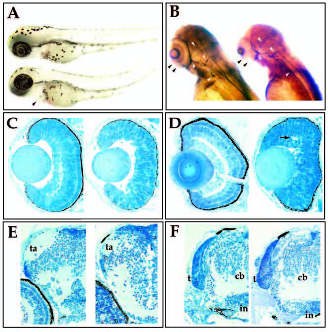
Fig. 1 Morphological characterization of the young mutation. (A) Lateral view of wild-type (upper) and young (lower) sibling embryos at 60 hpf. Mutant embryos are characterized by swelling of the pericardium (arrowhead) and a slight curvature of the tail. (B) 3A10 immunoreactivity in wild-type (left) and young (right) siblings at 72 hpf. Mutant embryos lack retinal plexiform staining (black arrowheads), but contain 3A10- positive axonal tracts in the brain and spinal cord (white arrowheads). (C) Transverse sections of 50 hpf wild-type (left) and young (right) retina. (D) Transverse sections of 72 hpf wild-type (left) and young (right) retina. Visible lamination is absent in the young retina, although small patches of plexiform regions can form (arrow). (E) Transverse sections through the forebrain of wild-type (left) and young (right) embryos at 96 hpf. Tectal arborization fields (ta) form in both. (F) Transverse sections through the hindbrain of wild-type (left) and young (right) embryos at 96 hpf. The tectum (t), cerebellum (cb), infundibulum (in) have formed in both the mutant and in wildtype fish.