
|
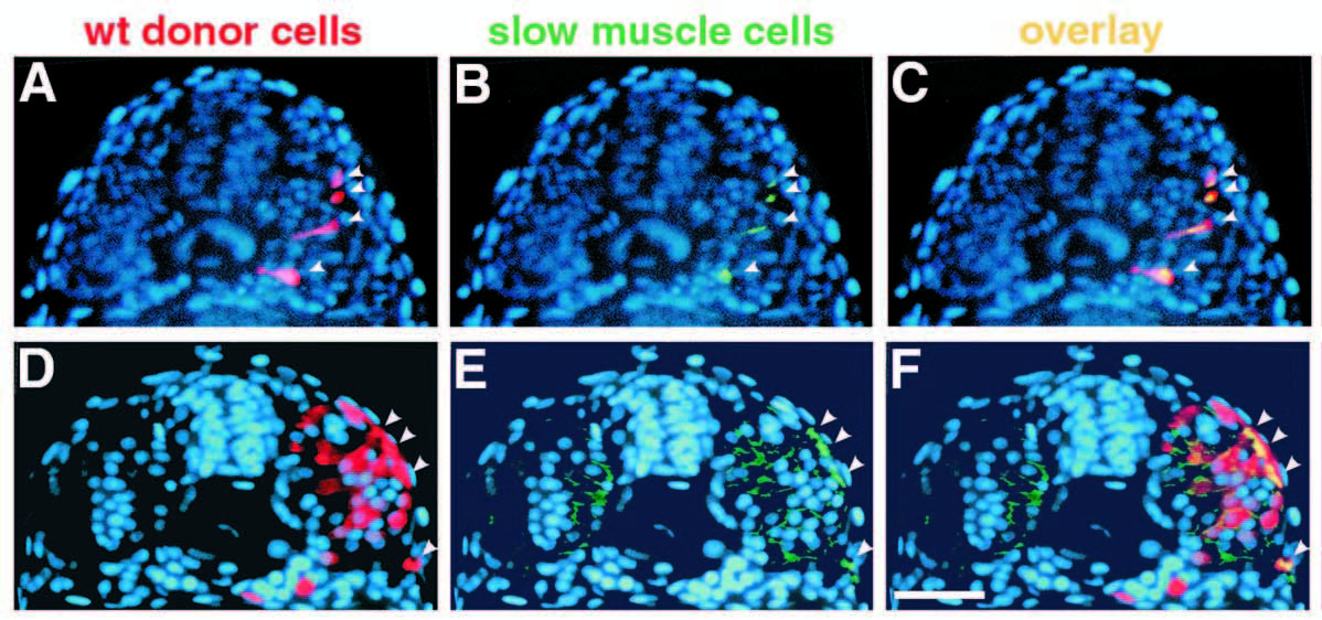
Fig. 5 Transplanted wild-type muscle cells rescue slow muscle development in smu-/-. Transverse sections from two different smu-/- embryos show donor-derived, wild-type muscle cells. Sections were labeled with the F59 antibody, detected with a fluorescein-conjugated secondary antibody, and counterstained with Hoechst 33258 (blue). (A,D) Rhodamine-labeled wild-type cells (red, arrowheads). (B,E) F59-labeled slow muscle fibers (green, arrowheads). (C,F) merged micrographs. In A-C, four transplanted wild-type cells have developed into slow muscle fibers in an approx. 22h smu-/- host (C, yellow, arrowheads). Cells are still migrating through the somite. In D-F, transplanted wild-type cells have differentiated into both slow (4 cells; F, yellow, arrowheads) and fast (approx. 10 cells) muscle fibers in a 24h smu-/- host. Slow fibers can be distinguished from fast on the basis of the intensity of F59 labeling. Bar, 50 μm.