
|
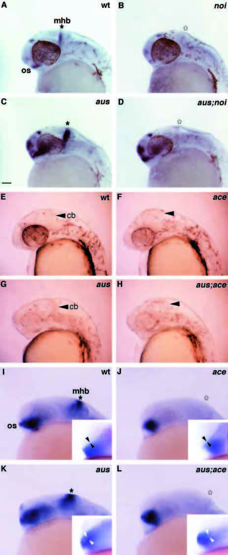
Fig. 9 aus is unlikely to represent a mutation in the ace or noi genes. Lateral views of prim-12 (A-H) and prim-5 stage (I-L) embryos with rostral to the left. Asterisks indicate the position of the mhb. (A-D) Analysis of ace expression in wild-type (A), noi (B), aus (C) and aus;noi double mutant (D) embryos. ace expression is absent at the mhb in the absence of functional Noi and is upregulated in the forebrain both in aus and aus;noi double mutant embryos. (E-H) Appearance of wild-type (E), ace (F), aus (G) and aus;ace double mutant (H) embryos. In the aus;ace double mutant, the rostral brain looks similar to the aus phenotype while the absence of cerebellum (arrowheads) is characteristic of the ace phenotype. (I-L) Analysis of noi expression in wild-type (I), ace (J), aus (K) and aus;ace double mutant (L) embryos. noi expression is absent at the mhb in the absence of functional Ace. The aus dependent upregulation of noi in the eyes and forebrain (K) is much reduced in the absence of functional Ace (L). The inset panels in I-L show the width of the optic recess (arrowheads) used as a landmark to infer the genotype the embryos – phenotypic morphological differences are much more visible in living embryos prior to the in situ protocol (see E-H). Abbreviations: cb, cerebellum; mhb, midbrain/hindbrain boundary; os, optic stalk. Scale bar: 50 μm.