
|
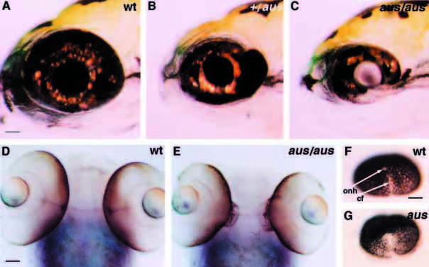
Fig. 4 Differentiation of the eyes is perturbed in aus mutant embryos. Lateral (A-C) and ventral (D,E) views of the eyes. (A-C) In heterozygous aus mutant embryos at early larval stage, there is ectopic outgrowth of the temporal retina (B) and, in putative homozygous aus mutant embryos, the ventral retina is reduced (C) as compared to wild-type siblings (A). (D,E) Ectopic outgrowth at the back of the retina is observed in the aus mutant embryo at early larvae stage (E) as compared to a wild-type sibling (D) (the faint blue staining is noi expression within the eyes, optic nerves and midline). (F,G) In the putative homozygous aus mutant embryo at prim-20 stage (G) there is incomplete closure of the optic fissure as compared to a wild-type sibling (F). See also Fig. 6G,H. Abbreviations: cf, choroid fissure; onh, optic nerve head; wt, wild type. Scale bars: (A-C) 50 mm; (D-G) 25 μm.