
|
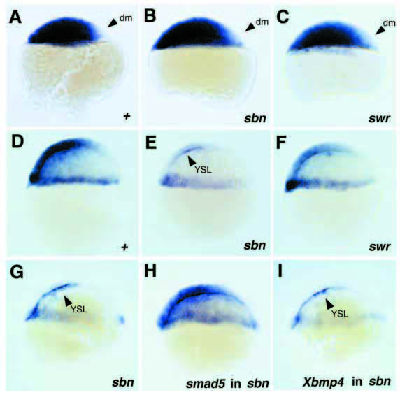
Fig. 5 Expression of bmp2b in sbn and swr mutant embryos (A-F) and after injection of smad5 or Xbmp4 mRNA in sbn mutants (G-I). All embryos are shown in a lateral view, dorsal side right. (A,D) Wild-type siblings of swr mutants; (B,E) sbn mutants from cross of sbn heterozygous female with wild-type male. Embryos were fixed and stained in parallel to the swr and wild-type embryos shown; (C,F) swr mutants. (A-C) Sphere stage; presumptive dorsal mesoderm (dm) devoid of bmp2b staining is marked with arrowheads. (D-F) Shield stage; the sbn mutant (E) displays severely reduced bmp2b staining, and the swr mutant (F) slightly reduced bmp2b staining in the blastoderm; the staining in the yolk syncytial layer (YSL, arrowhead in E) appears normal. (G-I) sbn mutant embryos, shield stage, after injection of smad5 (H; 50 pg/embryo) or Xbmp4 (I; 4 pg/embryo) mRNA. Similar results as in I were obtained for approx. 100 embryos in each of three independent injection experiments. Of the Xbmp4-injected sbn sibling embryos that were raised to day 2, 53% (n=45) showed a strong morphological response, appearing wild-type or ventralized (see Fig. 6).