
|
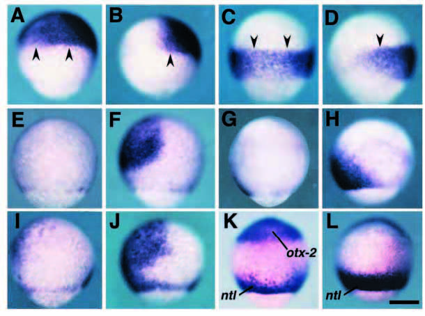
Fig. 6 DN-BRIA causes the expansion of the neuroectoderm to the ventral part without any perturbation of the anterior-posterior pattern. At the 80% epiboly stage, embryos injected with mRNA encoding DN-BRIA (A,C,E,G,I,K) and b-galactosidase (B,D,F,H,J,L) were stained with the specific probes for otx-2 (A,B), hoxa-1 (C,D), gata-3 (E,F), eve-1 (G,H) and zbmp-2 (I,J). For the embryos shown in K and L, a mixture of probes for otx-2 and ntl was used. (A-J) Embryos are oriented with their dorsal side to the right and animal pole to the top; (K,L) ventral views with the animal pole at the top. In embryos injected with DN-BRIA mRNA, expression domains of anterior (otx-2, A) and posterior (hoxa-1, C) neuroectoderm marker genes expanded to the ventral part of the embryo, compared with the negative control embryos (B,D, stained with probes for otx-2 and hoxa-1, respectively). The posterior boundary of otx-2 and anterior boundary of hoxa-1 were maintained (arrowheads) in the DN-BRIA-injected embryos. By contrast, ventral nonneural ectodermal marker genes, gata-3 and eve-1, are reduced by the overexpression of DN-BRIA (E and G, respectively). Similarly, zbmp-2 is also reduced (I). This suggests that the neuralization of the ectoderm (A,C) and reduction of non-neural ectoderm (E,G) presented here results from the down-regulation of zbmp-2, caused by interference with the autoactivation process by DN-BRIA. (K,L) DN-BRIA cannot induce involuting dorsal mesoderm as revealed by the axis-like expression pattern of ntl even in the embryo in which neuroectoderm is expanded to the most ventral part. Scale bar, 200 μm.