
|
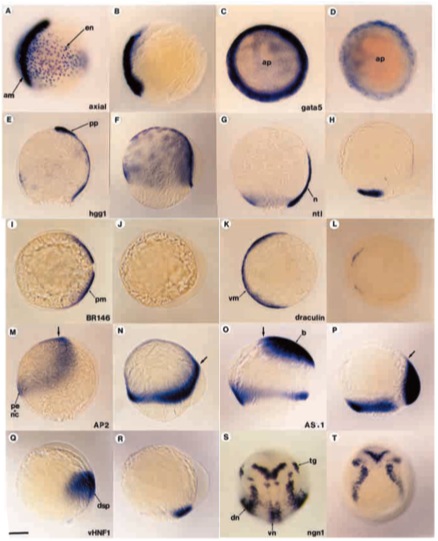
Fig. 4 Antivin inhibits mesoderm formation at gastrulation. Embryos injected with antivin RNA were analyzed by in situ hybridization with different molecular probes. For each probe, the wild-type sibling and the corresponding injected embryo are shown next to each other (left and right, respectively). (A,B) Labelling with axial and (C,D) labelling with gata5 for low doses of antivin RNA (1 pg or 0.5 pg, respectively) showing the deletion (A,B) or strong reduction (C,D) of endoderm (en) during gastrulation. (E,F) Labelling with hgg1, a marker of anterior prechordal plate (pp) and yolk syncytial layer. (F) Overstaining with hgg1 reveals the absence of prechordal plate in atv-injected embryos. (G,H) Absence of axial mesoderm is revealed using ntl, which labels the notochord (n) as well as the margin. (I,J) Absence of paraxial mesoderm (pm) is detected probing with BR146. (K,L) draculin, a marker of ventral mesoderm (vm) reveals that atv-injected embryos lack this territory. (M,N) Expression of AP2, specific to presumptive epidermis (pe) and neural crest (nc), is enlarged and shifted dorsovegetally (arrow). (O,P) The presumptive brain (b), labelled with AS11, is located at the margin rather than at the animal pole. (Q,R) Expression of vHNF1, which marks the presumptive dorsal spinal cord (dsp), is shifted to the lateral margin. (S,T) Expression of neurogenin1 (ngn1) reveals the presence of proneural clusters except in the ventral midline. Arrows show the anterior tip of the embryonic axis. dn, dorsal proneural clusters; tg, trigeminal ganglion; vn, ventral proneural clusters. Scale bar: 150 μm.