
|
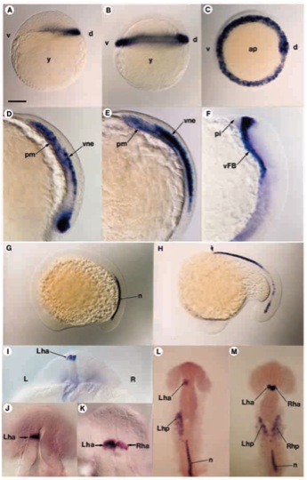
Fig. 2 Distribution of antivin transcripts during embryogenesis. (A) atv transcripts are first detected at the sphere stage at the dorsal margin of the embryo. (B,C) 40% epiboly. In lateral view in B and polar view in C, antivin RNA is localized all around the margin. (D) At gastrulation, atv is expressed in the prechordal mesendoderm (pm) and in the overlying ventral neurectoderm (vne). (E) As the mesoderm expressing atv migrates anteriorly, labelling increases in the neurectodermal layer. (F) At the onset of somitogenesis, atv expression is located in the ventral forebrain (vFB) territory. (G) At the 10-somite stage, atv starts to be expressed in the caudal part of the notochord (n). (H) By the 16-somite stage, anterior notochord cells (arrow) express atv, while transcripts disappear posteriorly. (I) At the 20-somite stage, optical cross-section of the diencephalon showing atv expression in the left habenula nucleus (Lha). (J) In 92.8% embryos, atv is expressed in the left part of the habenula. (K) In 4.5% embryos, both left and right sides of the habenula are labelled. (L) When located in the left part of the habenula, atv is also expressed in the left heart primordium (Lhp). (M) When located on both sides of the habenula, atv is also seen in both left and right heart primordia. ap, animal pole; d, dorsal; L, left; pi, pillow; R, right; Rha, right habenula; Rhp, right heart primordium; v, ventral; y, yolk. Scale bar, 150 μm (A,B,D,E,G,H,J,K), 300μm (C,F,I,L), 75μm (M-Q).