
Image
|
Figure Caption
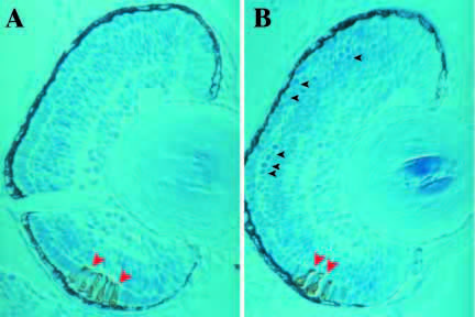
Fig. 7 Frontal sections through retinae at 72 hpf stained with mAB Fret43, which stains photoreceptors after their final mitosis. (A) Wild-type retina is already layered into the major three layers and photoreceptors begin appearing in the ventral patch (red arrowheads). (B) nrf/nrf mutant also shows differentiating red/green cones (red arrowheads) but, in addition in the mutant, multiple rounded cells, not integrated into the tissue are distinguished (small black arrowheads).
Figure Data
Acknowledgments
This image is the copyrighted work of the attributed author or publisher, and
ZFIN has permission only to display this image to its users.
Additional permissions should be obtained from the applicable author or publisher of the image.
Full text @ Development