
Image
|
Figure Caption
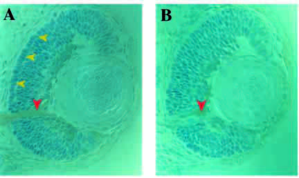
Fig. 6 Cross sections through retinae at 50 hpf prepared from whole mounts stained with monoclonal antibody Zn5. (A) Wild type; note the Zn5-positive retinal ganglion cells and the optic nerve (red arrowhead). In the back of the retina, a layer of prospective photoreceptor cells can be distinguished (yellow arrowheads). (B) nrf/nrf sibling, note that Zn5-positive cells and optic nerve are present (red arrowhead), but that layering of the retina is not as apparent.
Figure Data
Acknowledgments
This image is the copyrighted work of the attributed author or publisher, and
ZFIN has permission only to display this image to its users.
Additional permissions should be obtained from the applicable author or publisher of the image.
Full text @ Development