
|
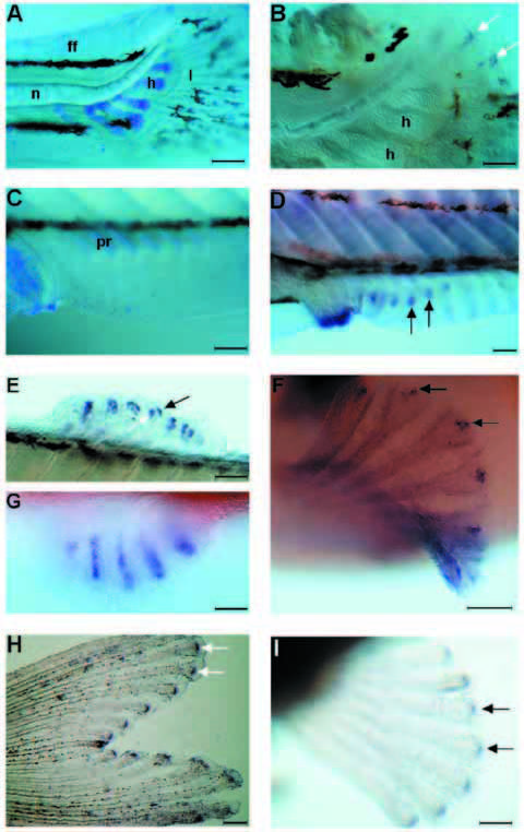
Fig. 3 Shh and ptc1 expression in the developing fins of zebrafish larvae. (A,B) Caudal fins of 5.5 mm larvae. (C,D) Anal fins of 6.5 mm larvae. (E) dorsal fin, (F) pectoral fin and (G) pelvic fin of 7.2 mm larva. (A) Alcian blue staining of the cartilage. The lepidotrichia stained in light blue are already well developed in the caudal fin fold. (B) Caudal part of a similar larva to the one in A showing shh expression in each developing lepidotrichia. Note that whole-mount in situ hybridization procedure causes a shrinkage of the tissues. (C) Alcian blue staining of the cartilage of the anal fin. Some elements of the endoskeleton as well as the exoskeletal lepidotrichia are not yet apparent. (D) In a comparable fin to that in C, shh transcripts are already found in the fin fold in subset of cells where the lepidotrichia are forming. Shh is also strongly expressed in the analia-genitalia region. (E-G) Shh expression subsequently appears in dorsal (E), pectoral (F), and pelvic (G) fin rays. The waving of the dorsal fin (E) shows the two groups of cells, one per side, expressing shh. Ptc1 expression in a caudal fin (H) and a pectoral fin (I) of a 7 mm larva. The caudal fin is already heavily pigmented. Arrows in B,D-F and in H,I indicate shh and ptc1 expression, respectively. Anterior is to the left; h, cartilage condensation of hypural bones; l, lepidotrichia; n, notochord; ff, fin fold; pr, proximal radial of the endoskeleton of an anal fin. Scale bars, A, 200 μm; B, 100 μm; C,D,E,G,H,I, 160 μm; F, 80 μm