
Image
|
Figure Caption
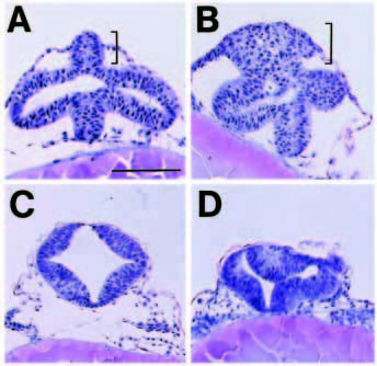
Fig. 6 The dorsal neural tube expands in six3 mRNA-injected embryos. Transverse sections of embryos injected with globin (A,C) or six3 mRNA (B,D). (A,B) Six-somite stage; sections through the prospective forebrain and optical vesicles. Dorsal (bracket) but not ventral region contains extra cells in six3 mRNA-injected embryos. The size of cells is basically unchanged. (C,D) 24 hours; Sections through the midbrain. Structure of dorsal portion of the neural tube is disorganized, but cell number is not changed substantially. Scale bar, 100 μm.
Acknowledgments
This image is the copyrighted work of the attributed author or publisher, and
ZFIN has permission only to display this image to its users.
Additional permissions should be obtained from the applicable author or publisher of the image.
Full text @ Development