
|
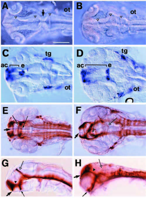
Fig. 5 Disorganized head formation in six3 mRNA-injected zebrafish embryos. 20 pg of globin (A,C,E,G) or six3 mRNA (B,D,F,H) were injected into 2-cell-stage zebrafish embryos; see Fig. 7 for quantitative results. (A,B) Dorsal views at 24 hours. Three ventricles (open triangles) in the brain are filled with masses of cells and the midbrain-hindbrain boundary (arrow) is disorganized in six3 mRNA-injected embryos. (C,D) Dorsal views of Islet-1 expression at 24 hours. The region between AC and epiphysis (bracket) is enlarged in six3 mRNA-injected embryos. Likewise, the distance between left and right trigeminal ganglia is expanded. (E-H) Dorsal (E,F) or lateral views (G,H) of 36 hour embryos labeled with antibody against acetylated α-tubulin. AC (arrow), POC (thin arrow), SOT (black triangle) and region surrounded by them are enlarged, while axons in PC (open arrow) are reduced in six3 mRNA-injected embryos. Additional abbreviations: ot, otic vesicle; tg, trigeminal ganglion. Scale bar, 200 μm.