
|
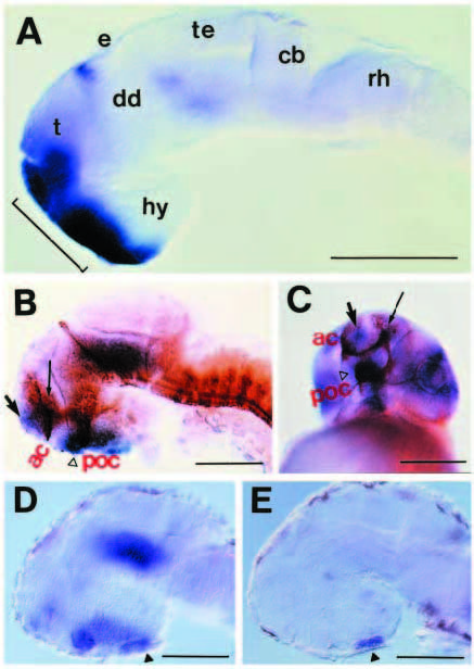
Fig. 4 six3 expression in later development. (A) Lateral view of 24- hour embryo showing six3 expression at the rostral surface of the forebrain (bracket); the eyes were removed. (B,C) Lateral (B) or frontal (C) view of six3 expression at 36 hours. The six3 signal (blue) is localized in the medial telencephalon (arrow), optic stalk (open triangle) and near the olfactory nerve (thin arrow). Axons were stained with antibody against acetylated α-tubulin (brown). (D,E) Lateral views of six3 (D) or lim3 (E) expression at 48 hours. six3 is expressed in the pituitary anlage (black triangle). Additional abbreviations: ac, anterior commissure; cb, cerebellum; dd, dorsal diencephalon; e, epiphysis; hy, hypothalamus; poc, postoptic commissure; rh, rhombomeres; t, telencephalon; te, tectum. Scale bars, 200 μm.