
Image
|
Figure Caption
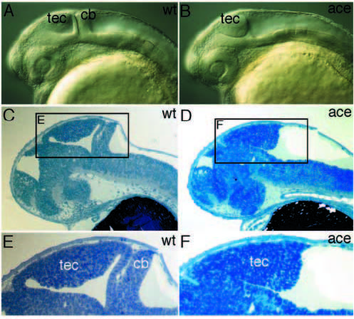
Fig. 7 Brain phenotype of acerebellar embryos. (A,B) At pharyngula stage, mutant embryos lack a cerebellum and the midhindbrain fold, but show an enlarged tectum (lateral view of living embryos). (C,D) Sagittal section of 36 hour embryos. (E,F) High magnification view of area depicted in C and D, showing the midhindbrain phenotype in more detail. cb, cerebellum; tec, tectum.
Figure Data
Acknowledgments
This image is the copyrighted work of the attributed author or publisher, and
ZFIN has permission only to display this image to its users.
Additional permissions should be obtained from the applicable author or publisher of the image.
Full text @ Development