
IMAGE
Fig. 6
- ID
- ZDB-IMAGE-140305-59
- Publication
- Appel et al., 1998 - Regulation of neuronal specification in the zebrafish spinal cord by Delta function
- All Figures
- Figures for Appel et al., 1998
Image
|
Figure Caption
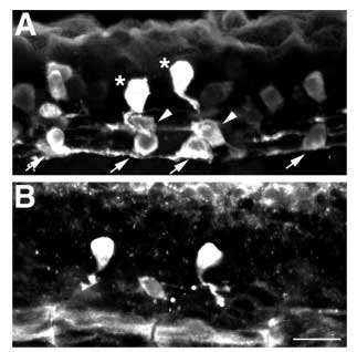
Fig. 6 Ventral interneurons are reduced in the absence of lateral inhibition. (A) Lateral view of 24 h spinal cord showing distribution of GABA-positive neurons. KA interneurons (arrows) differentiate in ventral spinal cord, VeLD interneurons (arrowheads) are slightly more dorsal and CoSA interneurons (asterisks) are dorsal to VeLDs and just ventral to RBs. (B) Lateral view of a 24 h embryo injected with X-Delta-1STU and nlacZ mRNAs and labeled with anti-GABA. A single VeLD and two CoSA interneurons have differentiated. Scale bar, 10 μm.
Acknowledgments
This image is the copyrighted work of the attributed author or publisher, and
ZFIN has permission only to display this image to its users.
Additional permissions should be obtained from the applicable author or publisher of the image.
Full text @ Development