
Fig. 2
- ID
- ZDB-IMAGE-140305-2
- Publication
- Pack et al., 1996 - Mutations affecting development of zebrafish digestive organs
- All Figures
- Figures for Pack et al., 1996
|
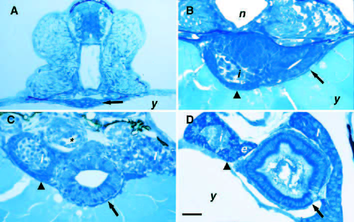
Fig. 2 Histology of the digestive organs in the developing zebrafish. Cross sections of proximal gut. (A) A small lumen is visible in the developing intestine (arrow) of the 36 hour postfertilization (hpf) embryo. (B) The intestine has enlarged (arrow) and the pancreatic islet (i) and exocrine precursor cells (arrowhead) are visible in the 52 hpf embryo. (C) The intestinal epithelium (arrow) has begun to polarize in the 72 hpf larvae. Exocrine precursors surround the islet (arrowhead). (D) The intestinal epithelium (arrow) has fully polarized and the pancreatic exocrine cells have differentiated (arrowhead) at 4 dpf. The liver is rostral to the plane of section in A and B. e, exocrine pancreas; i, pancreatic islet; l, liver; n, notochord; y, yolk; * labels pneumatic duct of gas bladder. Scale bars, A and D, 25 μm; B, 20 μm; C, 30 μm.