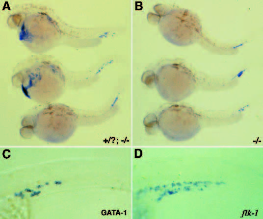
Fig. 7 GATA-1 is expressed in clo mutant embryos only in the region outlined by the flk-1-expressing cells. GATA-1 expression in wild-type and clo mutant embryos at 30 hpf. (A) Lateral view of 2 wild-type (top) and one clo mutant (bottom) embryos. (B) clo mutant embryos. (C,D) Higher magnification view of cells expressing GATA-1 (C) and flk-1 (D) in the lower trunk and tail regions of clo mutant embryos. GATA-1 expression is not detected at early stages in clo mutant embryos. Expression appears as flk-1 is being detected and only in the region lined by the flk-1-positive cells. Additional analysis reveals that although GATA-2 does not seem to be expressed in clo hematopoietic tissues, the GATA-1- expressing cells appear to mature normally as assessed by positive diaminofluorene staining used to detect heme (data not included).
Image
Figure Caption
Acknowledgments
This image is the copyrighted work of the attributed author or publisher, and
ZFIN has permission only to display this image to its users.
Additional permissions should be obtained from the applicable author or publisher of the image.
Full text @ Development

