
|
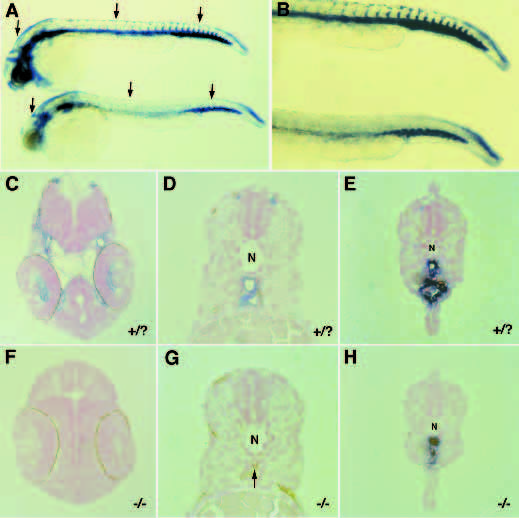
Fig. 5 Histological examination of wild-type and clo mutant embryos overstained with flk-1 (see Methods). Lateral view of the 36 hpf embryos used for histology (A,B), wild-type above and mutant below; transverse sections of the head, trunk and tail regions of wildtype (C,D,E) and clo mutants (F,G,H) respectively. Arrows in A indicate the A-P position of the sections shown. (A,B) flk-1 expression is seen in the lower trunk and tail regions of mutant embryos but is missing from the rest of the trunk. In the head region, flk-1 expression is observed in intracranial vessels in wild-type (C) but not in mutants (F); there is also staining on the dorsal aspect of the hindbrain region which is seen in both wild-type and mutant embryos (and can be observed at earlier stages (Fig. 2A-C)), as well as staining on the ventrolateral aspects of the caudal head region which appears on sections as hazy background on the outside of overstained embryos (data not included). (D,G) In the the trunk region, while vessels are clearly stained in wild-type embryos, no flk- 1 expression is seen in mutants and in fact, somitic tissue appears to occupy the space where the dorsal aorta and axial vein usually form (G, arrow). (E,H) In the tail region, flk-1-expressing cells are present in both wild-type and clo mutant embryos although at an apparently reduced number in the mutants. N, notochord.