
|
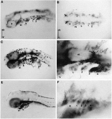
Fig. 5 Late expression pattern of snail1 in the head. Embryos were dissected and the yolk was removed. (A) 24 hour embryo, dorsolateral view and (B) 24 hour embryo, dorsal view. snail1 RNA is localized in various cells around the eyes (e) and otic capsule (ot), and in pharyngeal arch primordia (mandibular arch (m), hyoid arch (h), and the first (1) and second (2) gill segments). D, Dorsal; V, ventral; R, right; L, left. (C) 36 hour embryo, lateral view. The extent of labelling has expanded in the branchial arches and is easily distinguishable in the mandibular (m) arch and the hyoid (h) arch, and in the first four (1, 2, 3 and 4) gill arches. Pectoral fin buds (pf) also accumulate snail1 transcript. (D) High magnification of the ear region (ot, otic capsule) and caudal branchial arches focusing on snail1 RNA expression in gill arches. The mesenchyme of each arch is composed of neural crest cells and paraxial mesodermal cells. (E) 60 hour embryo, lateral view. snail1 RNA disappears from the middle of the arches where chondrocytes are beginning to differentiate. (F) Details of the embryo shown in E, focusing on branchial arches.