
|
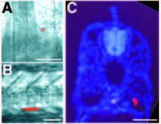
Fig. 3 Lateral presomitic cells become deep muscle cells. (A) Dorsal view of the segmental plate of an approximately 15 h embryo (12 somites), after injection of a lateral presomitic cell with lysinated rhodamine dextran. (B) Side view of the same embryo at about 40 h. The lateral presomitic cell developed into two ventral muscle cells located in somite 16. (C) Transverse section of the same embryo, counter stained with Hoechst 33258 to show cell nuclei. Both cells are deep muscle fibers. In a series of similar experiments, 25 out of 25 injected lateral presomitic cells became deep muscle fibers. In side views, anterior is to the left; in dorsal views, anterior is up; in transverse sections, dorsal is up. Scale bars, 50 μm.