
Fig. 2
- ID
- ZDB-IMAGE-140228-25
- Genes
- Antibodies
- Publication
- Schilling et al., 1996 - The chinless mutation and neural crest cell interactions in zebrafish jaw development
- All Figures
- Figures for Schilling et al., 1996
|
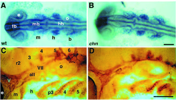
Fig. 2 Early neural crest development and the formation of sensory neurons appear unaffected in chn. (A,B) 15 hour, dorsal view. Wholemounted embryos showing dlx2 expression in three major streams of migrating neural crest cells that form the arches. All embryos from a cross between two identified heterozygous chn/+ parents show identical patterns of dlx2 expression. (C,D) 30 hour, lateral view. Pharyngeal arches at prim-15 stage labeled in situ with the monoclonal antibody, zn-5 (Trevarrow et al., 1990). The antibody labels the cell surfaces of segmental clusters of neurons in hindbrain rhombomeres, sensory neurons of the cranial ganglia and endodermal pouches that form between pharyngeal arch primordia. Abbreviations: all, anterior lateral line; b, branchial arches; e, eye; fb, forebrain; h, hyoid arch; hb, hindbrain; m, mandibular arch; mb, midbrain; o, otic vesicle; p3-6, pharyngeal arches 3-6; r2-6, rhombomeres 2-6; V, trigeminal ganglion; VII, facial ganglion. Scale bars, 200 μm.