
|
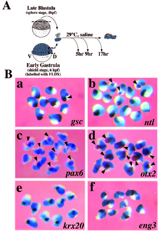
Fig. 4 Anterior neural markers are induced in animal caps following conjugation to shields. (A) Schematic representation of cap/shield conjugate preparation and culture. Each conjugate consisted of five animal caps from sphere stage embryos (late blastula, 4 hpf) fused to a single shield from a shield stage embryo (early gastrula, 6 hpf) and placed in culture (see Material and Methods). Conjugates were harvested when control embryos reached the end of gastrulation (10 hpf, 5 hours in culture), the 12 somite stage (15 hpf, 9 hours in culture) or the end of somitogenesis (24 hpf, 17 hours in culture). D, dorsal; V, ventral. (B) Whole-mount in situ hybridization analysis of mesodermal and neural gene expression in cap/shield conjugates. Conjugates were analyzed for gsc (a) and ntl (b) expression after 5 hours in culture, for pax6 (c), otx2 (d) and krx20 (e) expression after 9 hours in culture and for eng3 (f) expression after 17 hours in culture. The shield lineage label (see Materials and Methods) is visualized in light blue and the animal cap portion is unlabelled. Induction of gene expression in the animal cap portion of the conjugates is indicated by arrowheads. This data is included in Table 2.