
|
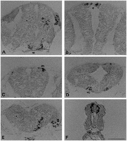
Fig. 3 Expression pattern of Isl-1 at 22 hpf. (A) Cross section through the anterior part of the brain in front of the eyes; (B) cross section through the anterior brain at the level of the epiphysis; (C) cross section at the level of the nucleus of posterior commissure; (D) cross section of the midbrain; (E) cross section of the 4th segment of the hindbrain; (F) cross section through the spinal cord of a double stained (anti-tubulin and anti-Isl-1) embryo. ag, acoustic ganglion; vr, ventrorostral cluster; dr, dorsorostral cluster; ob, olfactory bulb; pc, nucleus of posterior commissure; vc, ventrocaudal cluster. Bar, 50 μm.