
|
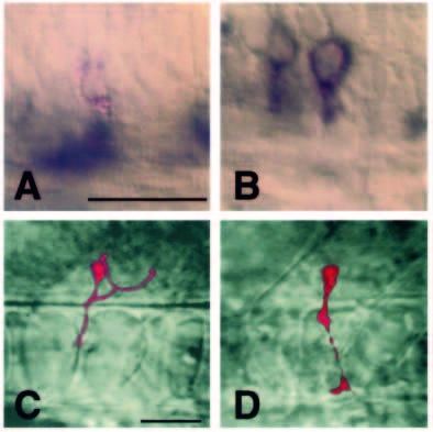
Fig. 7 islet2 gene expression in transplanted primary motoneurons determined by in situ RNA hybridization. (A,B) Cells transplanted from the MiP position to the CaP position at different developmental stages and fixed and probed for islet2 expression after the transplanted cells developed axons. Transplanted cells are marked by red staining and islet2 localization by blue. Scale bar in A equals 25 mm for A,B. Scale bar in C equals 25 μm for C,D. (A) This cell was transplanted about 1 hour before axogenesis. It maintained the MiP fate and did not express islet2. Hybridization signal in the ventral spinal cord represents late islet2 expression, probably in secondary motoneurons. (B) This cell was transplanted 2-3 hours before axogenesis, adopted a CaP fate, and expressed islet2. (C) Image of the cell in A prior to fixation showing its MiP axonal projection. (D) Image of the cell in B prior to fixation showing its CaP axonal projection.