
Image
|
Figure Caption
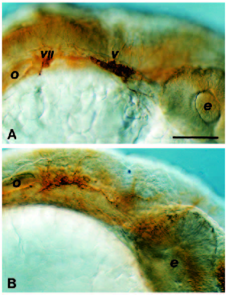
Fig. 9 The preotic cranial ganglia are abnormally formed in Hoxa-1- injected embryos. This is seen by comparing 24 hour embryos stained with anti-acetylated tubulin to reveal the VIIth and Vth ganglia between the otic vesicle (o) and the eye (e). In control embryos (A), these ganglia are distinct but, in embryos injected with Hoxa-1 (B), they are reduced and fused into one ganglion immediately anterior to the otic vesicle. Scale bar, 100 μm.
Acknowledgments
This image is the copyrighted work of the attributed author or publisher, and
ZFIN has permission only to display this image to its users.
Additional permissions should be obtained from the applicable author or publisher of the image.
Full text @ Development