
|
Fig. 4
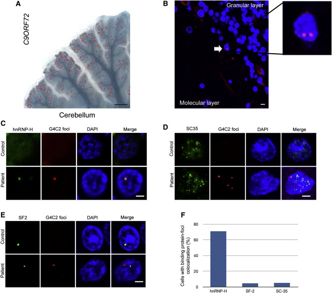
Intranuclear Neuronal RNA Foci in C9ORF72 Mutant ALS and FTD Brain Tissues Colocalize Very Closely with hnRNP-H
(A) Image of mutant C9ORF72 patient cerebellum overlaid with the location of neurons containing G4C2 RNA foci (red dots) (scale bar represents 4 mm).
(B) G4C2 RNA foci-positive neurons (white arrow) were observed between the granular and molecular layer of the cerebellum (scale bar represents 10 μm).
(C-E) FISH and ICC were performed for hnRNP-H (C), SC35 (D), and SF2 (E), with a G4C2 mutation-negative ALS case used as control (scale bar represents 3 µm).
(F) The percentage of foci that colocalized with hnRNP-H, SF2, and SC-35 were counted (n = 50 cells). Of the three RNA binding proteins that colocalized with foci in transfected cells, only hnRNP-H shows a striking degree of overlap for 70% of all foci in the cerebellum.
See also Figure S4.