
|
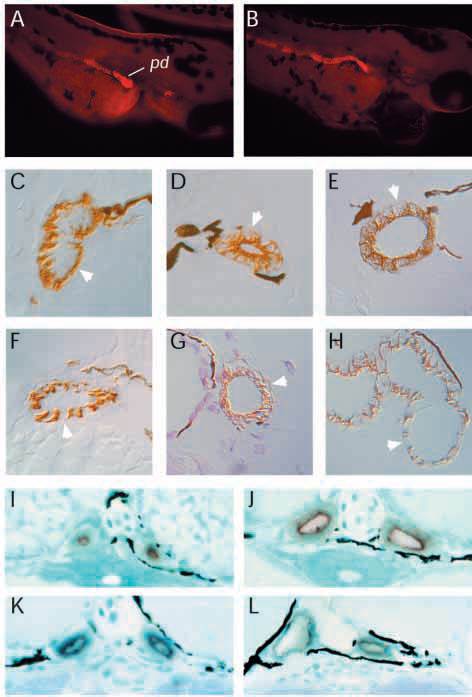
Fig. 10 Altered membrane protein targeting in pronephric mutants. (A) The monoclonal antibody alpha6F raised against the chicken alpha-1 subunit of the Na+/K+ ATPase reacts strongly with the pronephric duct (pd) at hatching (2.5 days) in wild-type (A) and mutant (dbbm468) (B) whole-mount embryos. In sections of wild-type duct (C), alpha6F staining is exclusively basolateral (arrow) while, in dbb (D), alpha6F staining is strongly apical and diminished on basolateral membranes (arrow). Diminished alpha6F basolateral staining is also observed in (E) fleer (flr), (F) dizzy (dzz), (G) inflated (ifl) and (H) big league chew (chw) mutant duct cells. (A,B) Cy3- labeled secondary antibody; (C-H) HRP-labeled secondary antibody. (I-L) Endogenous alkaline phosphatase staining is apical in wild-type pronephric duct (I) and is not affected in dbb (J), inflated (ifl) (K), or dizzy (dzz) (L), mutant duct cells.