
|
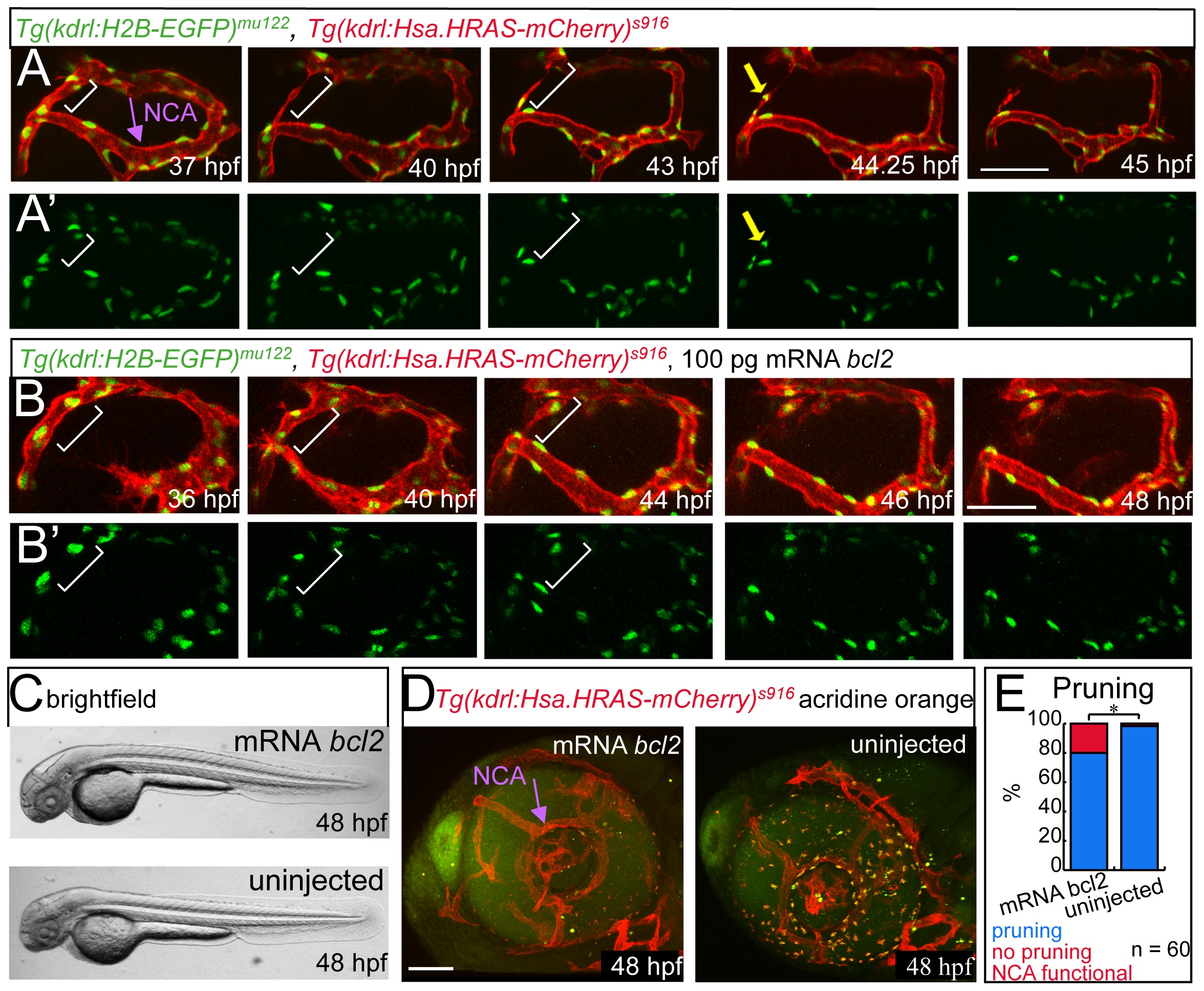
Fig. 3
Endothelial cell apoptosis during CrDI pruning.
All images in lateral view with anterior to the left. (A) Stills of time-lapse imaging of Tg(kdrl:H2B-EGFP)mu122, Tg(kdrl:Hsa.HRAS-mCherry)s916 from 37-45 hpf. (A′) Tg(kdrl:H2B-EGFP)mu122 channel only. White brackets indicate dorsal CrDI. Yellow arrow in (A) and (A′) at 42.25 hpf time point points to dying cell. (B) Stills of time- lapse imaging of Tg(kdrl:H2B-EGFP)mu122; Tg(kdrl:Hsa.HRAS-mCherry)s916 embryos from 36-48 hpf injected with bcl2 RNA. No dying cells are detectable in the CrDI. (B′) Tg(kdrl:H2B-EGFP)mu122 channel only. (C) Brightfield images of bcl2 mRNA or uninjected embryos. (D) Acridine orange staining on bcl2 mRNA injected or control embryos. (E) Quantification of CrDI pruning at 48 hpf.