
|
Fig. 1
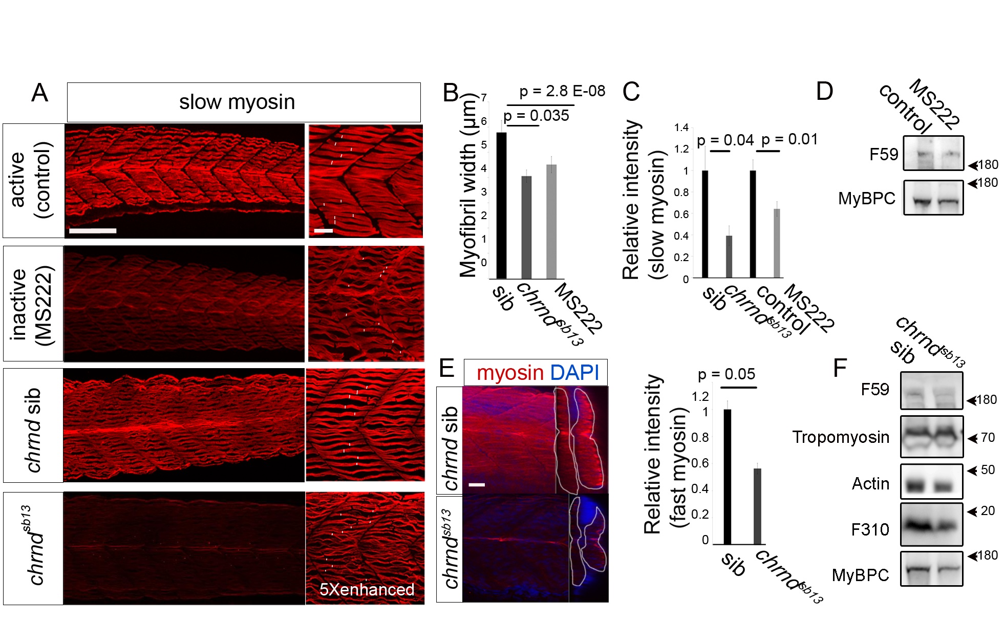
Muscle activity enhances zebrafish myofiber width and myosin level.
Muscle activity was blocked either by adding MS222 to the fish water for 24chrndsb13/sb13 mutants lacking the acetylcholine receptor delta subunit, which were identified by their immotility. (A) Confocal stacks of slow MyHC immunostaining in 48 hpf embryos. Note the reduced myofibril content and poor bundling in inactive fish. White bars in the right-hand panels indicate minimal myofibrillar bundle width on each fiber. Fivefold more laser light was used to generate the lower right image. (B) Width of myofibrillar material in well-bundled regions of >5 myofibers in somite 17 were measured from >6 embryos in each condition. (C) Slow MyHC level relative to control in n = 15 embryos. (D and F) Western analysis of 48 hpf mutant or MS222-treated embryos, compared to respective controls. (E) Confocal stacks of embryos stained for general MyHC (A4.1025) in lateral (left image) and transverse (right image, somite indicated by white line) view. Graph shows relative MyHC level. n = 10 embryos. Bars represent SEM and samples were compared by t test. All experiments were repeated at least twice. Scale bar = 90 μm in (A, left), (E), and 23 μm in (A, right).