
|
Fig. 3
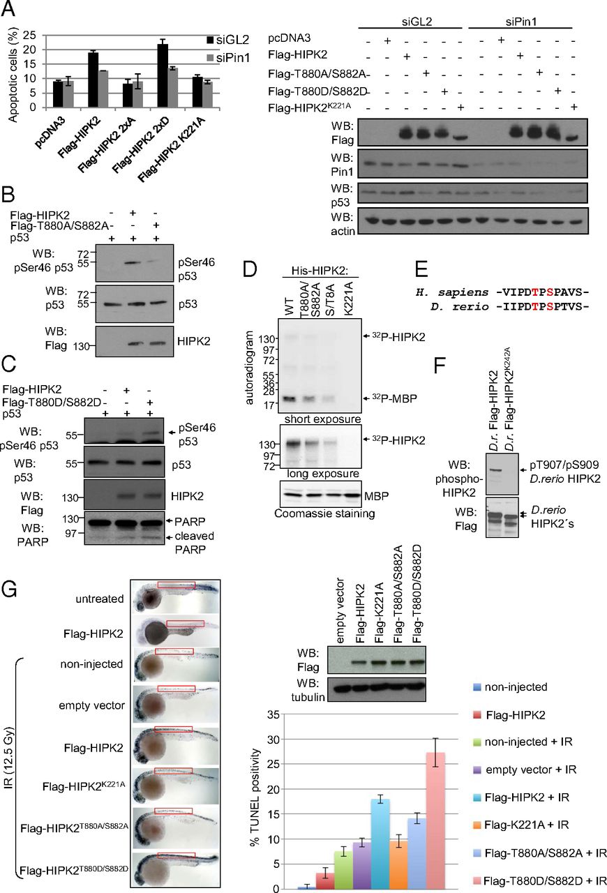
HIPK2 autophosphorylation at Thr880/Ser882 regulates its apoptotic function in human cells and zebrafish embryos. (A) HIPK2 autophosphorylation is critical for its apoptotic function. HCT116 cells were transfected with empty vectors and HIPK2 expression vectors as indicated. Cells were analyzed by FACS for apoptosis-associated annexin V-positivity after annexin V/propidium iodide staining. The graph shows means and SD. n = 3. siGL indicates control siRNA transfected cells (black bars); siPin1 indicates cells depleted of Pin1 (gray bars). Flag-HIPK2, Pin1, and p53 levels were determined by immunoblotting. A representative result is shown. (B and C) HIPK2 autophosphorylation at Thr880/Ser882 regulates p53 Ser46 phosphorylation capacity. H1299 cells were transfected with the expression vectors indicated, and cell lysates were analyzed by immunoblotting. (D) HIPK2 autophosphorylation correlates with its kinase activity. Bacterially expressed 6xHis-HIPK2 proteins were used for radioactive in vitro kinase assay with MBP as substrate and analyzed by autoradiography. Input was controlled by Coomassie brilliant blue staining and HIPK2 immunoblotting. Input levels of the His-HIPK2 proteins are shown in Fig. 1C, which was performed in parallel to this experiment here by using the same batch and amounts of His-HIPK2 proteins. (E) The HIPK2 autophosphorylation site is conserved between zebrafish and humans. (F) D. rerio HIPK2 is autophosphorylated. H1299 cells were transfected with expression vectors coding for wild-type or kinase-deficient zebrafish HIPK2. Total cell extracts were analyzed by immunoblotting using the indicated antibodies. (G) HIPK2 autophosphorylation at T880/S882 potentiates apoptosis induction in zebrafish embryos. Embyos were microinjected with mRNAs coding for HIPK2 proteins as indicated, exposed to IR (12.5 Gy), and analyzed and quantified for TUNEL positivity.