
|
Fig. 3
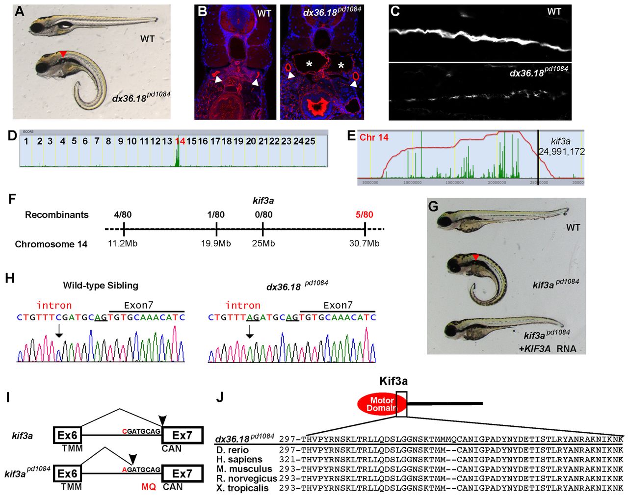
kif3apd1084 mutants present defects in cilia formation and kidney cysts. (A) Brightfield image of 5-dpf WT sibling and dx36.181084 mutant larvae. Red arrowhead indicates a kidney cyst. (B) Confocal images of transverse sections of 5-dpf WT sibling and dx36.181084 mutant larvae stained with phalloidin (red) and DAPI (blue). The arrowheads point to the pronephric ducts and the asterisks mark kidney cysts. (C) Confocal image of 3-dpf WT and dx36.181084 mutant in whole-mount, stained for acetylated tubulin to mark cilia. (D) Linkage analysis for dx36.181084 using SNPtrack. (E) The homozygosity interval for dx36.181084. (F) Linkage analysis of the dx36.181084 locus. (G) Rescue of the dx36.18pd1084 phenotype with WT human KIF3A cRNA. Rescue was confirmed by genotyping (not shown). (H) Sequencing of genomic DNA of WT and kif3apd1084 mutant larvae showing a six nucleotide insertion in kif3a that adds two extra codons between exons 6 and 7 (underlined). (I) Sequencing of cDNA confirms the insertion of codons coding for MQ in kif3apd1084 mutants. TMM, threonine, methionine, methionine; CAN, cysteine, alanine, asparagine. (J) Alignment of Kif3a protein sequences from zebrafish, human, mouse, rat and frog.