
Fig. 5
|
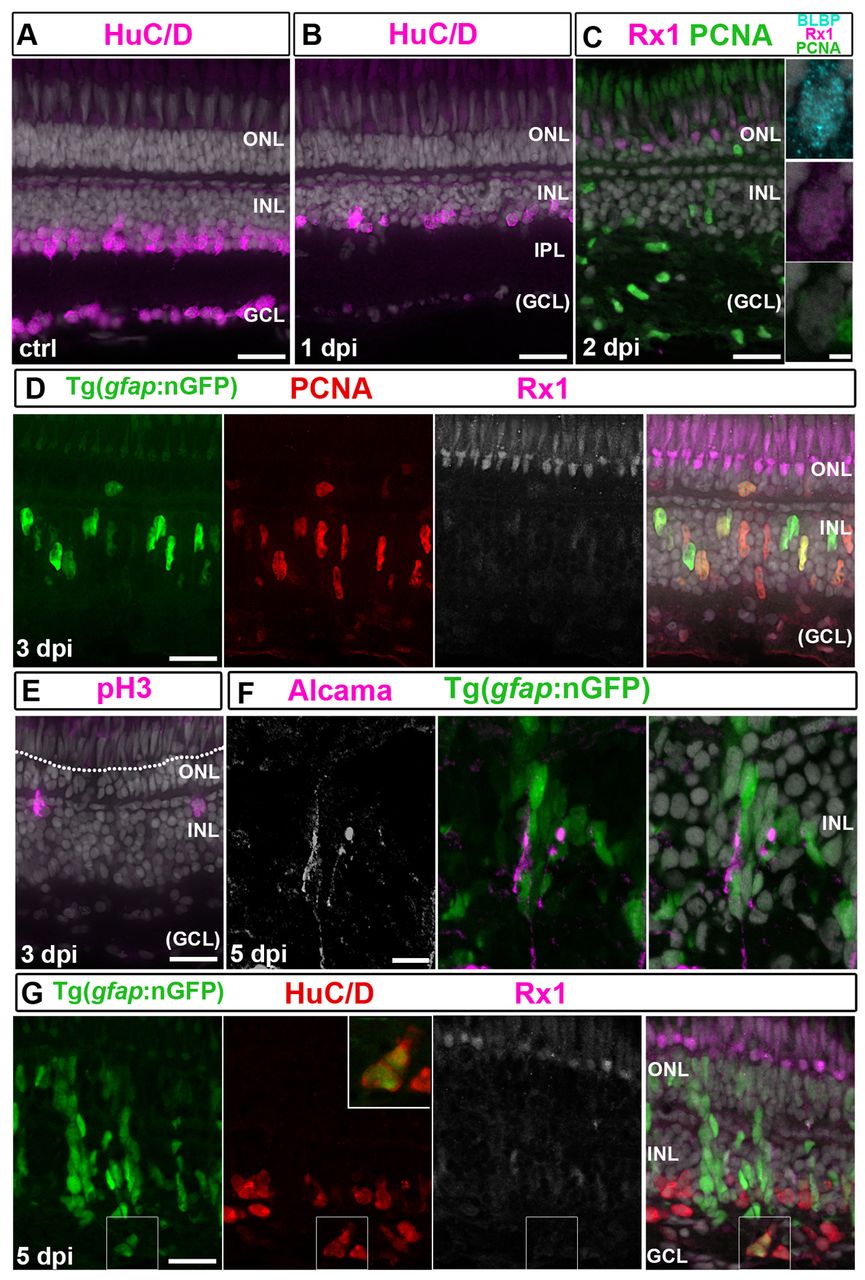
Fig. 5
Induction of neuroepithelial markers in Müller glia is delayed after ouabain. (A) Control (ctrl) HuC/D+ neurons. (B) HuC/D immunoreactivity is absent in the GCL and reduced in the INL at 1 dpi. (C) PCNA (green), BLBP (cyan) and Rx1 (magenta) staining at 2 dpi. Right panel: BLBP+/Rx1+ Müller glia. Scattered PCNA+/BLBP-/Rx1- cells in the INL and GCL are microglia. (D) At 3 dpi, nGFP+/PCNA+ (red)/Rx1+ (white/magenta) Müller glial nuclei are apically displaced. (E) At 3 dpi, pH3+ nuclei (magenta) are apical in the INL but not at the OLM (dotted line). (F) Alcama+ (white/magenta) Müller glia radial processes at 5 dpi. (G) At 5 dpi, nGFP+/HuC/D+ (red) regenerated neurons (higher magnification of boxed area shown in inset). Scale bars: 20 μm (A-G); 2 μm (inset in C).