
Image
|
Figure Caption
Fig. S9
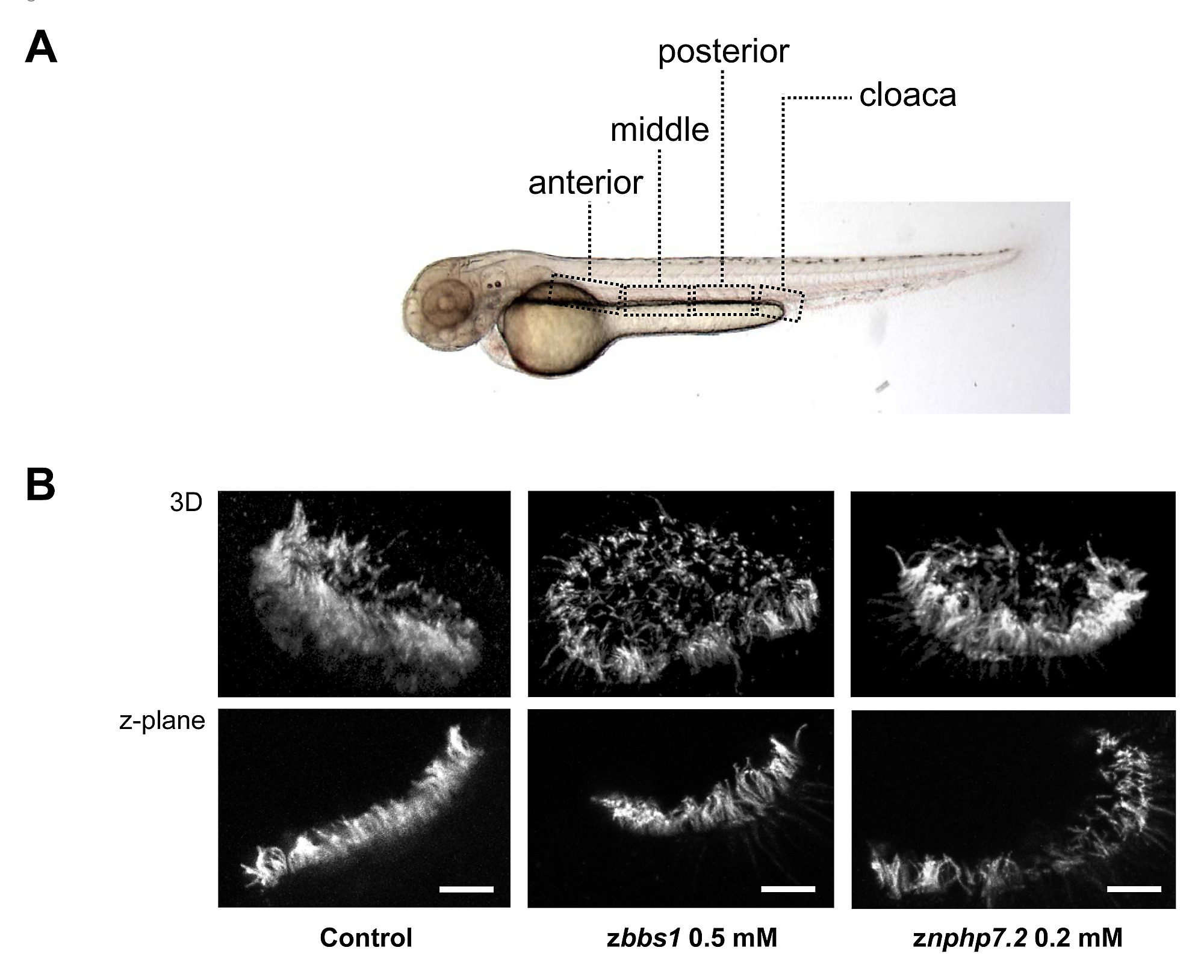
The morphants of zbbs1 and znphp7.2 exhibited impaired motility of cilia in the pronephric tubule. (A) The areas where the movies were recorded are shown by the dashed box. (B) Acetylated tubulin staining demonstrated normal development of cilia in the nasal pit (Scale bar = 10 µm). 3-dimensional (3D) images (upper panel) and z-plane images (lower panel) show that the cilia formation in the morphants of zbbs1 and znphp7.2 is normal compared to control embryo.
Acknowledgments
This image is the copyrighted work of the attributed author or publisher, and
ZFIN has permission only to display this image to its users.
Additional permissions should be obtained from the applicable author or publisher of the image.
Full text @ PLoS One